Sickle Cell Retinopathy

Image by Wikimedia Commons
https://youtube.com/shorts/0YxzDNCima0?feature=share

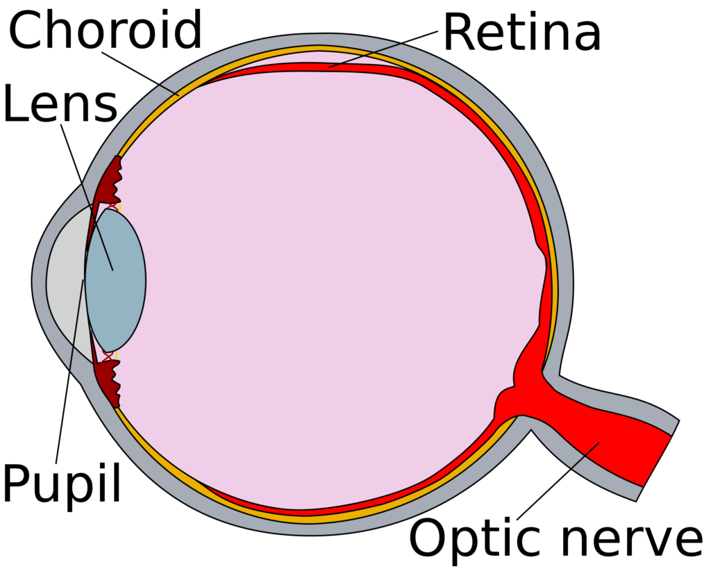
Sickle cell retinopathy is the separation of the retina and choroid within in eye, due to blockages in blood vessels caused by sickle red blood cells.

Retinopathy in sickle cell disease is caused by vessel occlusion and/or decreased blood flow due to increased viscosity – increased number of red blood cells – in the vessels of the retina.  This vessel blockage leads to lowered levels of oxygen (hypoxia) and the increase of small vessels that are very unstable and not attached to the retina. These new vessels (neovascularization) are weaker and can rupture, causing bleeding into the eye.1  This can lead to the retina separating (detachment) and blindness. 

These developments are painless and can go unnoticed by the patient unless bleeding (red blood cells) obscures the macula, which is where vision takes place.  The signs of retinopathy are “floaters” which are red cells that can be seen in the visual field, areas of decreased vision seen as black spots or blurry vision.  

Children rarely complain of these findings and retinopathy can begin at a young age. It is recommended that all children have a dilated eye examination starting at the age of 10 years and annually thereafter.

Over 40% of people who have hemoglobin SC will develop retinopathy in their lifetime and about 15% of people who have SS will develop retinopathy in their lifetime.

Retinopathy Study

A twenty-year study of retinopathy conducted in Jamaica with sickle cell disease patients revealed that this retinopathy was five times more common in hemoglobin SC disease compared to SS disease.3

By the age of about 25 years, 43% of people with SC disease had retinopathy compared to 14% with SS.  There is a possibility of regression (there was some improvement) of this disease, about 16% and a possibility of progression (becoming worse), about 14%.  Visual loss was uncommon (1 of 82), though these individuals were only followed until age 26 years.  Twenty years before this study a smaller study of people untreated for retinopathy reported that progression was most common between 20 and 40 years of age and spontaneous regression was more common in hemoglobin SS disease.

Review

Retinopathy in Children with Sickle Cell Disease

In children about 12% will be diagnosed with sickle cell retinopathy.  11% with nonproliferative retinopathy and about 2% with proliferative retinopathy.  Of this group 21% had hemoglobin SC and 9% had hemoglobin SS in the non-proliferative group and 5% versus 1% in the proliferative group.  The youngest in the non-proliferative group was about five years old in the SC group versus 6 years in the SS group. In the proliferative group the ages of onset were 12 years and 15 years respectively.  The recommendations for proliferative retinopathy was to begin screening at 9 years for SC and 13 years for SS.4

Proliferative retinopathy is what is described with sea fanning, bleeding into the eye.  Non-proliferative retinopathy is defined as other vascular changes (salmon patches, black sunburst spots, etc.)

Retinopathy in Sickle Cell Trait and C Trait (Hemoglobin AS and AC)

Retinopathy has been reported in both hemoglobin AS and AC (sickle cell trait and hemoglobin C trait).  These are case reports of one to several cases and in all of the reports the patients also had other diseases that contribute to retinal disease such as diabetes mellitus and hypertension.5,6  It can’t be said that either S or C traits are susceptible to retinopathy from these anecdotal reports.

Resources

  1. Jee K, Rodrigues M, Kashiwabuchi F, et al. Expression of the angiogenic mediator, angiopoietin-like 4, in the eyes of patients with proliferative sickle retinopathy. PLoS One 2017;12(8):e0183320. DOI: 10.1371/journal.pone.0183320.
  2. Ali MA, Shalchi Z. Sea-fan neovascularization in sickle-cell retinopathy. QJM 2021;114(10):747-748. DOI: 10.1093/qjmed/hcab174.
  3. Downes SM, Hambleton IR, Chuang EL, Lois N, Serjeant GR, Bird AC. Incidence and natural history of proliferative sickle cell retinopathy: observations from a cohort study. Ophthalmology 2005;112(11):1869-75. (http://www.ncbi.nlm.nih.gov/entrez/query.fcgi?cmd=Retrieve&db=PubMed&dopt=Citation&list_uids=16171867; http://www.aaojournal.org/article/S0161-6420(05)00889-4/abstract; https://www.aaojournal.org/article/S0161-6420(05)00889-4/fulltext).
  4. Li J, Bender L, Shaffer J, Cohen D, Ying GS, Binenbaum G. Prevalence and Onset of Pediatric Sickle Cell Retinopathy. Ophthalmology 2019;126(7):1000-1006. DOI: 10.1016/j.ophtha.2019.02.023.
  5. Hingorani M, Bentley CR, Jackson H, et al. Retinopathy in haemoglobin C trait. Eye 1996;10 ( Pt 3):338-42. (http://www.ncbi.nlm.nih.gov/entrez/query.fcgi?cmd=Retrieve&db=PubMed&dopt=Citation&list_uids=8796159; http://www.nature.com/eye/journal/v10/n3/pdf/eye199670a.pdf).
  6. Jackson H, Bentley CR, Hingorani M, Atkinson P, Aclimandos WA, Thompson GM. Sickle retinopathy in patients with sickle trait. Eye 1995;9 ( Pt 5):589-93. (http://www.ncbi.nlm.nih.gov/entrez/query.fcgi?cmd=Retrieve&db=PubMed&dopt=Citation&list_uids=8543078; http://www.nature.com/eye/journal/v9/n5/pdf/eye1995145a.pdf).