Hemoglobin

Why is blood red? What carries oxygen around the body? The answer to all these questions is hemoglobin. That tells us quite a lot about the importance of hemoglobin in our blood.

Red blood cells are mostly made up of hemoglobin. Hemoglobin binds oxygen in the lungs, and releases oxygen all around the tissues of your body, delivering it to your muscles and vital organs.

If we have an abnormal hemoglobin structure, it can make red cells less efficient at transporting oxygen.

A low level of hemoglobin is called anemia. Hemoglobin is measured by a complete blood count (CBC) test which is a routine blood test. Hemoglobin levels differ between people. For example, females tend to have lower levels than males, Dehydration can also cause hemoglobin levels to rise temporarily.

History

It’s 1840, and a German scientist called F.L. Hünefeld is studying an earthworm (of all things) under a microscope. He notices rectangular, bright-red structures in the earthworm’s blood – he’s just accidentally discovered hemoglobin – though there wasn’t a name for it yet. These crystals were later named as “Haemoglobin” by Hoppe-Seyler in 1864. Around 1870, Claude Bernard discovered its role as oxygen carrier. In time, another scientist by the name of Claude Bernard will later discover that the function of hemoglobin is to transport oxygen.

Fast forward to 1935, when American Biochemist Linus Pauling published his research on the structure of hemoglobin. Pauling found out how hemoglobin changes its structure when it binds with oxygen, and he discovered the molecular basis of sickle cell anemia. Though he contributed to our understanding of hemoglobin, many of Paulig’s views were highly unethical.

In 1962, molecular biologist Max Putz along with Sir John Kendrew won the Nobel Prize for Chemistry after discovering the detailed three-dimensional structure of hemoglobin by X-ray crystallography.

Structure

Hemoglobin is made up of four globin molecules. There are two alpha globin molecules and two beta globin molecules in each hemoglobin molecule. Each of the globins contains a heme, the receptor for oxygen.

The interesting thing about heme is that it changes shape when bound to oxygen. This change in the heme changes the shape of the whole hemoglobin molecule. Without oxygen the hemoglobin molecule is said to be in the “tense” state (T state). When one oxygen molecule binds to it, it makes it more likely that a second molecule will also bind. Then the molecule changes to the “relaxed” state (R state). In its relaxed state, it has a much greater affinity for the next two oxygen molecules. This occurs in the lungs when the hemoglobin in the red cell is exposed to oxygen. In the tissue, carbon dioxide attaches to the hemoglobin molecules and the oxygen is released.

Types of Hemoglobin

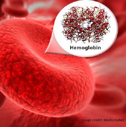
There are three main types of hemoglobin – embryonic, fetal, and adult.

  • Embryonic hemoglobin, There are four embryonic hemoglobin molecules that are made during embryonic development. The last to form is fetal hemoglobin.
  • Fetal hemoglobin is present in the fetus and in babies. Fetal hemoglobin binds to oxygen better than adult hemoglobin as before birth the baby obtains oxygen from their mother. After birth there is what is called a “hemoglobin switch” and adult hemoglobin is made and fetal hemoglobin declines.
  • Adult hemoglobin usually replaces fetal hemoglobin at around the age of 6 months.

With the change to adult hemoglobin mutations in the alpha and beta globin molecules become apparent as these are the adult hemoglobin molecules. There are hundreds of hemoglobin mutations. Only a few cause disease. Some mutations lead to the absence of hemoglobin, these are the thalassemia mutations. Thalassemia mutations can be either in the beta or the alpha gene and can cause mild to severe disease depending on whether the gene is capable of making some hemoglobin or is unable to make any hemoglobin. In combination with hemoglobin S they cause sickle beta thalassemia. Alpha gene mutations can also occur in people who have sickle cell disease.

Other mutations change the hemoglobin itself these are the types of mutations that cause hemoglobin S and other types of hemoglobin that in combination with hemoglobin S that cause sickle cell disease. These structurally changed hemoglobin molecules are called “variants”. The common variant hemoglobin molecules are: hemoglobin C, hemoglobin D, and thalassemia mutations. There are other rare forms of sickle cell disease caused by other rare variants.
this is how sickle cell disease comes about. Sickle cell disease can be caused by multiple variants of hemoglobin, including Hemoglobin S, Hemoglobin C, Hemoglobin E, etc.

Hemoglobin S is the main hemoglobin variant in sickle cell disease. The mutation is in one of the beta subunits. The imbalance between alpha and beta subunits is called thalassemia. When the beta chain gene fails, it is called beta-thalassemia, because the type of thalassemia is determined by what molecule is abnormal. Mild thalassemia may present with few symptoms, while severe thalassemia can be life-threatening.

Sickle Cell Disease

The most common form of sickle cell disease is hemoglobin SSWith both inherited beta globin molecules being S.. The abnormal hemoglobin causes the red blood cells to take on a sickle shape, become weak, and die easily. Due to sickle cells, the body struggles to continuously create new red blood cells, resulting in fatigue. The sickle cells can also block blood vessels, causing complications such as pain crises and damage to tissues.

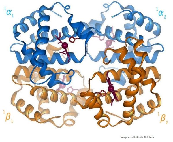
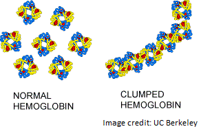
So, what causes these abnormal hemoglobin changes? Sickle cell is a genetic disease, meaning it is inherited from your parents. Hemoglobin is a protein and like all proteins it has a DNA code. This code is translated through RNA to amino acids that together form proteins within the cells. Sickle cell is a single mutation in the beta hemoglobin gene that changes this code for only one amino acid in the long chain that makes the beta globin protein. This single small change changes the properties of beta globin to allow polymerization leading to all of the problems with sickle cell disease.

Fetal hemoglobin therapy

Fetal hemoglobin (HbF) attracts oxygen better than normal adult hemoglobin (it has a higher affinity for oxygen) etal hemoglobin does not polymerize with hemoglobin S. There are genetic mutations that increase fetal hemoglobin in some people (hereditary persistence of fetal hemoglobin) who have hemoglobin S and they have no symptoms. It is estimated that 20% fetal hemoglobin would be protective against sickle cell disease. In adults, a therapeutic option for sickle cell disease is to reactivate fetal hemoglobin. Research has shown that even a small increase in HbF is sufficient to reduce symptoms of sickle cell disease.

One way to increase HbF in the body is through the drug, hydroxyurea. Another method of increasing HbF is through gene therapy. There are proteins (i.e. BCL11A) that have been shown to stop fetal hemoglobin production. Silencing these proteins can reactivate fetal hemoglobin production.

References